
The cell cycle is the series of steps by which living cells grow, duplicate their DNA, and divide into two identical daughter cells, each receiving one copy of the doubled material. The cycle is complete when each daughter cell is sealed in its own membrane, ready to start the cycle again.
The cycle occurs in a precisely orchestrated fashion to ensure cells pass a precise copy of their DNA on to their daughter cells. Errors at any point in the process — in the duplication of DNA prior to division or in the migration of DNA into the newly formed cells — can introduce errors into cells’ genetic programming that lead to cancer or other diseases.
Stages of the cell cycle
In cells with nuclei, the cycle occurs in five stages:
- G0, in which the cell isn’t dividing but performs its normal work in the body
- G1 (or gap1), in which the cell grows
- S (or synthesis), in which it copies its DNA
- G2 (or gap2), in which it prepares to divide
- M (or mitosis), in which the cell’s nucleus and cytoplasm (inner fluid) divide to form two daughter cells.
Human cells spend about 90% of their time in the first three stages, which are collectively known as interphase.
The duration of the cell cycle varies from one cell type to another. Most human cells complete the cycle in about 24 hours. Fast-growing cells, like those in the lining of the intestine, may complete it in just 9 or 10 hours, while liver cells take more than a year and neuronal cells take many years.
The cell cycle and cancer
Slip-ups or errors in any stage of the cell cycle — especially during the replication of DNA or allocation of DNA to the daughter cells — can set cells on a course for cancer.
The cycle includes several checkpoints, which function like stop-whistles on an assembly line, allowing the cell to scan for problems and take corrective action if necessary. Cells use these opportunities to make sure they’ve grown to the right size, have copied their DNA accurately, and have lined up their chromosomes properly for division. Mistakes may be fixed or, if severe, may prompt the cell to sacrifice itself to avoid passing them on to its descendants.
The checkpoints are operated by a variety of proteins. One of the most important of these is p53, which goes into action primarily at the G1 checkpoint in the cell cycle. When a cell’s DNA is damaged, a protein activates p53, which halts the cell cycle and orders repairs or, in the case of irreparable damage, triggers the cell’s death. So critical is p53 to the perpetuation of healthy cells that it’s known as the guardian of the genome.
If p53 is missing, malfunctioning, or less active than normal, it may allow a cell with damaged DNA to enter the next stage of the cell cycle, increasing the odds that DNA misspellings will be passed on to the daughter cells. The accumulation of such errors over multiple cell cycles can be a step toward cancer. p53 is the most commonly mutated gene in human cancers.
How does chemotherapy affect the cell cycle?
Different types of chemotherapy drugs attack cancer cells at various stages of the cell cycle.
Some target a specific stage of the cycle. Antimetabolites and antifols, for example, target the S stage and interfere with the construction of the DNA molecule. Bleomycin, etoposide, and other agents target the G2 phase, break DNA apart; and drugs known as vinca alkaloids target the M phase, which prevent chromosomes from lining up properly for cell division.
Other drugs act against dividing cells regardless of the cell cycle. These include alkylating agents, which bind to DNA and prevent it from being duplicated, and intercalators, which warp the normal shape and structure of the DNA double helix.
A third category are active against cancer cells regardless of which stage of the cycle they’re in. These include corticosteroids and hormone antagonists, which bind to cell receptors, preventing the cell from receiving growth signals.
How does radiation therapy affect the cell cycle?
Radiation kills cells that are actively dividing — that are not in the G0 stage. Because cancer cells grow out of control, they may spend more time undergoing division than normal cells do, making them especially vulnerable to radiation therapy.
How do targeted therapies disrupt the cell cycle?
Targeted therapies are a class of agents that inhibit specific cell proteins involved in cancer. A variety of these agents, some approved as standard therapy, some being tested in clinical trials, take aim at proteins that play a key role in the cell cycle.
The proteins CDK4 and CDK6, for example, stand guard over a critical portion of the cell cycle — the transition from the G0 and G1 phases into the S phase. In some types of cancer, abnormalities in these proteins can push cell division into high gear. Drugs that interfere with these proteins, known as CDK4/6 inhibitors, can help arrest the cell cycle. In some cases, the cancer cells not only stop dividing but lose all power to cycle and grow, causing tumors to shrink. CDK4/6 inhibitors have been shown to be most effective in treating advanced estrogen receptor-positive and HER2-negative breast cancer, and have shown activity in liposarcoma, non-small cell lung cancer, mantle cell lymphoma, melanoma, and glioblastoma.
Another example of a cell cycle protein targeted by drug therapy is WEE1. It’s part of a quality-control mechanism that pauses the cycle between the G2 and M stages so genetic errors can be fixed before the cell divides. Cancer cells without a functioning p53 protein may have accumulated substantial DNA damage by the time they reach the M stage. Drugs that block WEE1 allow them cruise into the M stage with so damage that they can’t survive. In a recent clinical trial led by Dana-Farber’s Joyce Liu, MD, MPH, a WEE1-targeting drug caused tumors to shrink in nearly one-third of patients with a hard-to-treat form of uterine cancer.
About the Medical Reviewer
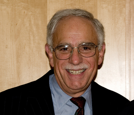
Dr. Mayer received his MD from Harvard Medical School in 1969, and postgraduate training in internal medicine at Mount Sinai Hospital, New York City. After fellowships in hematology and oncology at the National Cancer Institute and Dana-Farber Cancer Institute, he joined Dana-Farber in 1974.
In 1997, he was president of the American Society of Clinical Oncology. Dr. Mayer currently serves as Faculty Vice President for Academic Affairs at Dana-Farber Cancer Institute and Faculty Associate Dean for Admissions at Harvard Medical School.