The information used in diagnosing a brain tumor takes many forms. At Dana-Farber/Brigham and Women’s Cancer Center (DF/BWCC), patients’ brain tumor tissue undergoes a broad range of diagnostic tests: not only standard pathology exams in which tumor cells are viewed under a microscope, but also next-generation scans for mutated genes and misassembled chromosomes, as well as whole-genome searches for surplus or missing copies of genes.
Such extensive testing helps pinpoint the exact type and characteristics of a particular tumor. The more specific the diagnosis, the more precise the therapy can be.

But such a wealth of test results is only as valuable as pathologists’ and physicians’ ability to interpret it. As the diagnosis of brain tumors becomes more complex, DF/BWCC pathologists and cytogeneticists (specialists who focus on chromosome structure) routinely come together to consult one another, compare notes, and present physicians with a unified report on their diagnostic findings.
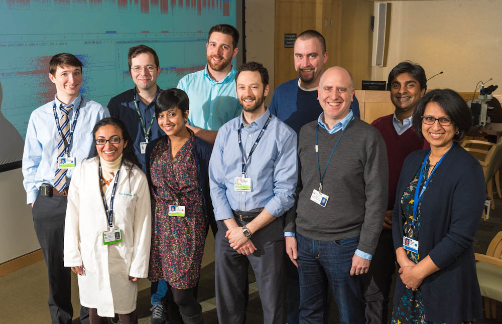
Keith Ligon, MD, PhD, along with his colleague (and wife) Azra Ligon, PhD, both of the Center for Molecular Oncologic Pathology (CMOP) at DF/BWCC, have established a brain tumor diagnostic tumor board, which draws specialists from across the various disciplines within pathology. At the meetings, attendees review individual brain tumor cases, presenting the results of the diagnostic tests within their specific area.
“The diagnosis of brain tumors today involves multiple streams of information from different tests,” says Keith Ligon, who is a principal investigator in the CMOP. “By bringing as much information as possible to bear on each patient’s case, we can ‘tighten’ the diagnosis – narrow it down as precisely as possible – reduce the chance of misdiagnosis, give really useful prognostic information, and help oncologists arrive at an effective treatment.”
Tumor tissue from brain cancer patients at DF/BWCC undergoes at least four categories of cellular and molecular tests. The first is a conventional histopathology test, in which the tissue is examined under a microscope for structural abnormalities, followed by an immunohistochemistry test, which focuses on cell proteins, called antigens, that signal a specific type of cancer. Then comes OncoCopy, a whole-genome scan that can detect extra or missing copies of certain genes. Another test, OncoPanel, examines the protein-coding portions of genes to identify mutations associated with cancer. (OncoPanel is part of the Profile program, directed by Neal Lindeman, MD, which analyzes tumor tissue for known cancer-related mutations.) All exams are ordered within two weeks of the tissue becoming available, with the hope that results will be available in four weeks.
Read more:
Every case of brain cancer at DF/BWCC is brought before the brain tumor diagnostic tumor board. “Each case generates several reports, reflecting the different type of testing done on the tissue,” Ligon explains. “At the diagnostic board meeting, we work to reconcile the results of these tests to produce an integrated pathology report that goes to the patient’s treating physician.
“Whenever new test results become available, we add them to the integrated report,” he continues. “Ultimately, we’d like to produce a report that would be available online to the physician and could be constantly updated.”
In some cases, this multi-test, collaborative approach has enabled DF/BWCC pathologists to change a diagnosis that had been made incorrectly at another institution, Ligon says.
One advantage of this comprehensive approach is that the test data, once collected, is available for future research. In a recent study, Shakti Ramkissoon, MD, PhD, Wenya Linda Bi, MD, PhD, Rameen Beroukhim, MD, PhD, and Brian Alexander, MD, MPH, used the data to show that the testing technologies are working well and suggested how they can be improved to capture even more data, Ligon notes.
“We can also store the tumor tissue and culture it to create a living tissue bank for patients,” Ligon adds. “This tissue can be implanted in mice to create avatars of patients’ tumors that are then used to study which treatments are likely to be most effective.”
