By Christopher Fletcher, MD, FRCPath
Chief of Onco-Pathology, Dana-Farber Cancer Institute
Vice Chair for Anatomic Pathology, Brigham and Women’s Hospital
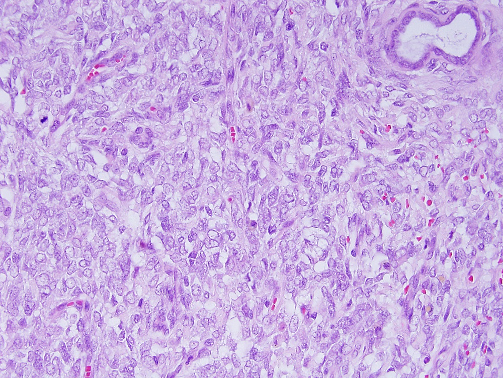
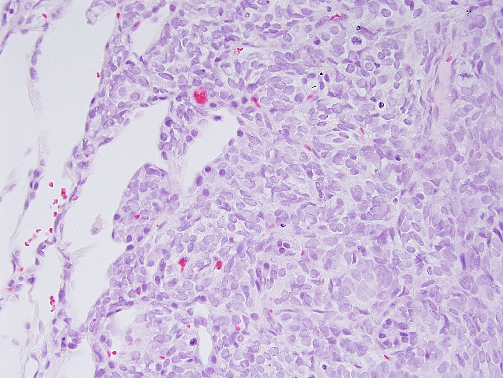
In order to determine whether a growth represents relapse of a previously diagnosed cancer or is a newly developed, separate tumor, doctors obtain a tissue sample from the patient and have it examined by a pathologist. The pathologist, who also examines the tissue to determine the grade and stage of the cancer, views the tissue under a microscope and compares it to tissue from the earlier cancer. If cells in the two samples are similar, it most often indicates the original tumor has relapsed or spread. (Federal regulations require that glass microscope slides and tissue blocks be retained for 10 years. For tumors that are more than 10 years old, direct comparison of the specimens may not be possible. However, at Brigham and Women’s Hospital (BWH), which serves as the Pathology Department for both BWH and Dana-Farber, we aim to retain materials indefinitely.)

Occasionally a patient may develop two separate cancers of the same type, which appear nearly identical under the microscope. This seems to occur most often in lung cancers, which can look very similar, making it difficult to separate a new primary tumor from metastasis of the original primary tumor, unless molecular testing is performed. In such cases, molecular genetic testing may be required to confirm these are two separate tumors, rather than a recurrence of a prior tumor.


is there an error? The pathologist is examining the tissue to determine the grade not the stage?
Hi Ronny,
We’ve made the correction — the pathologist examines the grade and the stage.
Best,
DFCI